Results
Video 1. Cell adhesion to the substrate. Can you guess which of these cells is ‘suffering’ from a lack of integrin αv?
Video 2. Fluorescently labeled EB3 protein located on the tips of growing microtubules allows monitoring of microtubule dynamics. Can you spot the difference?
Figure 1. The results of the analysis of the isolation of adhesion proteins by mass spectrometry show differences in the composition of focal adhesions and extracellular matrix proteins in cells in which the expression of αVβ5 integrin is altered
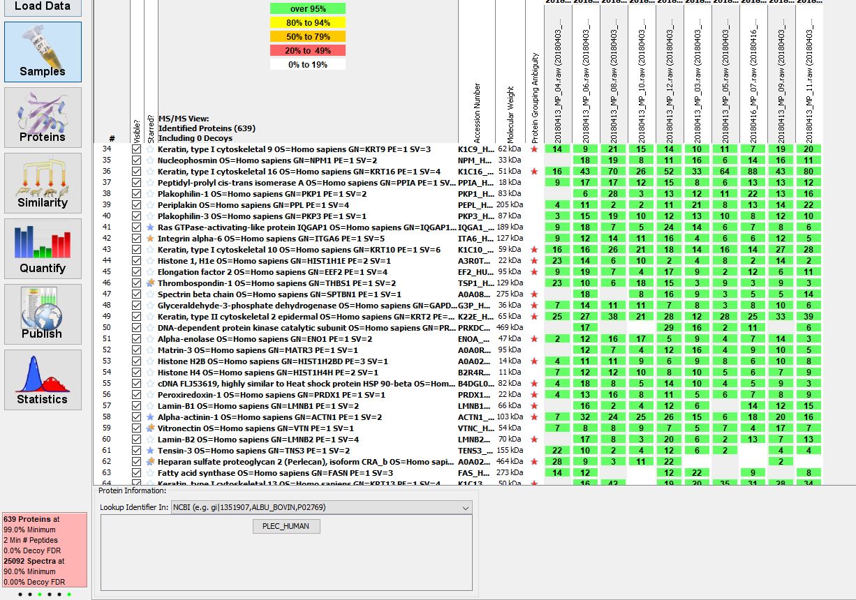
Figure 2. By changing the composition of focal adhesions by silencing the expression of individual proteins (talin 1 - red; talin 2 - green) we can affect the appearance of the cellular cytoskeleton (microtubules - purple; actin - yellow)
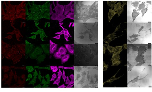
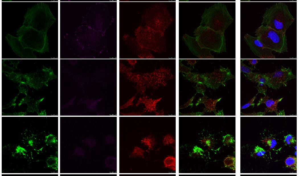
Figure 3. Cells in addition to focal adhesions (marker of focal adhesions vinculin - purple) located on the tips of stress threads of the actin cytoskeleton (green) also contain reticular adhesions (marker of reticular adhesions NUMB - red) located below the nucleus (blue)