

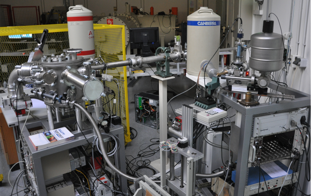
Linija za PIXE/RBS spektroskopiju
Spektroskopija emisije x-zraka inducirane česticama/protonima (engl. Particle/Proton Induced X-ray Emission, PIXE) je tehnika koja se koristi za određivanje elementarnog sastava uzorka izloženog snopu čestica. Kada se nabijene čestice (npr. protoni) energije u MeV-skom rasponu (obično između 2 i 3 MeV) kreću kroz materijal, one prvenstveno gube energiju pobuđivanjem elektrona u atomima materijala. Elektroni u unutarnjim ljuskama atoma (uglavnom K i L ljuske) dobivaju dovoljno energije da budu izbačeni. Elektroni iz vanjskih ljusaka popunjavaju ta prazna mjesta, što je praćeno emisijom x-zraka. Energije tih x-zraka su karakteristične za element i mogu se detektirati kako bi se utvrdio elementarni sastav uzorka. PIXE je relativno jednostavna i multielementarna analitička tehnika koja se može koristiti za mjerenje koncentracija elemenata u rasponu od natrija (Na) do urana (U). PIXE je također nedestruktivna tehnika, a zbog visokog omjera signala i pozadine, vrlo je osjetljiva za širok raspon mjerenih elemenata, s granicama detekcije blizu 1 ppm.
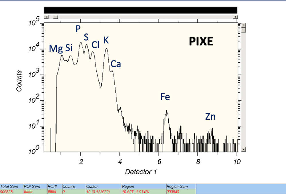
Primjer PIXE spektra.
Brzi ioni također mogu interagirati s atomskim jezgrama u uzorku putem elastičnih sudara. Tehnika nazvana Spektroskopija Rutherfordovog povratnog raspršenja (engl. Rutherford Backscattering Spectrometry, RBS) temelji se na detekciji povratno raspršenih iona (obično protona ili iona He i Li) pod kutovima blizu 180 stupnjeva. Mjerenjem energije i broja povratno raspršenih iona moguće je odrediti koncentraciju i dubinski profil elemenata u površinskim slojevima uzorka izloženog ionskom snopu. Ova je tehnika posebno moćna za dubinsko profiliranje teških elemenata u laganim supstratima. Kada se RBS izvodi u kombinaciji s PIXE-om, može se koristiti za određivanje koncentracija lakih elemenata, što samo s PIXE-om nije moguće.
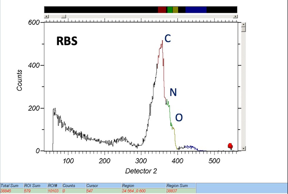
Primjer RBS spektra.
Tehničke specifikacije
Komora za rutinsku PIXE/RBS analizu instalirana je na –45° liniji Tandetron akceleratora. Opremljena je s dva PIXE detektora. SDD (engl. Silicon Drift Detector) detektor koristi se za analizu lakih elemenata (natrij i teži), dok Si(Li) detektor ima veliki prostorni kut i Mylar filtar optimiziran za detekciju teških elemenata (kalij, kalcij i teži). Komora ima integrirani izmjenjivač uzoraka koji može prihvatiti do 16 uzoraka (veličine između 10 i 25 mm). Uzorci su obično izloženi snopu protona od 2 MeV kružnog oblika (promjera 3, 5 ili 8 mm) i strujama snopa u rasponu od 1 do 10 pA.
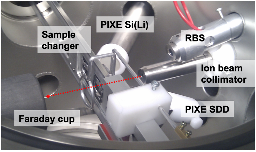
Pogled u komoru za PIXE/RBS spektroskopiju.
Prikupljanje i analiza podataka
U tipičnim radnim uvjetima, tri ADC-a (dva za PIXE i jedan za RBS) kontrolira se softverom za prikupljanje podataka SPECTOR. Prikupljeni PIXE spektri analiziraju se softverom GUPIX, dok se za analizu RBS spektara koristi softver SIMNRA.